


Common Non-Cancerous Breast Lumps
Although breast cancer is the most common cancer among women, the majority of breast lumps are not cancerous (benign).
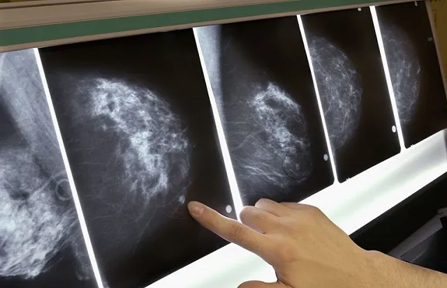
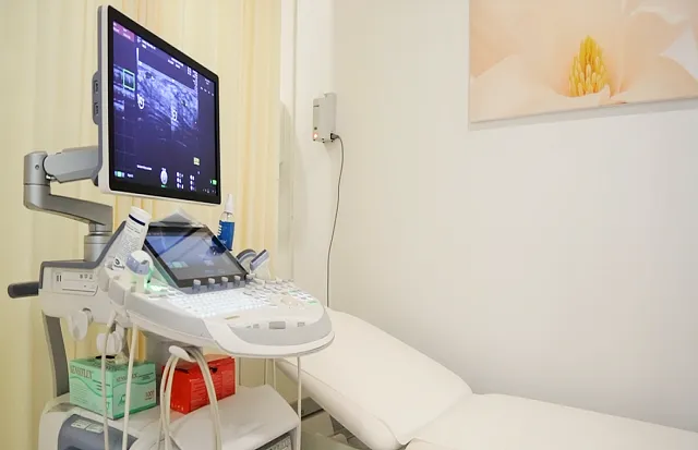
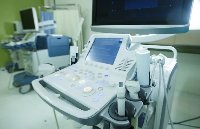
However, it can be difficult to differentiate a cancerous lump from a non-cancerous lump purely by a clinical examination. Therefore, if you find a breast lump, see your gynaecologist or GP. They should further refer you to a breast clinic for proper imaging, which will usually involve ultrasound, but for some also additional mammogram.
Breasts lumps can sometimes mimic changes found in breast cancer. In these cases it is necessary to confirm the diagnosis with a biopsy in order to make an accurate diagnosis and to differentiate between a benign breast condition and breast cancer.
Following are examples of common noncancerous (benign) breast lumps:
Fibroadenoma
Around 10-12% of women have a fibroadenoma, often without knowing about it. They usually develop between the age of 15-35, but can occur in women at any age. Although rare, they may also occur in men.
Fibroadenomas can be solitary or multiple, found in one or both breasts, palpable or impalpable. They can vary in size, and over time both increase or decrease in size or even sometimes disappear. It is not found in other organs than the breast tissue.
Symptoms:
- No associated symptoms
- Lumps in one or both breasts
- Tender or painful, particularly premenstrually
- Round or oval with distinct smooth borders
- Texture is firm, smooth or rubbery
- Easily movable under the skin
Causes:
Fibroadenomas develop from a lobule of the milk-producing gland. Surrounding glandular tissue and milk ducts grow over the lobule and form a solid lump. The cause for developing this is unknown, but it is believed to be due to increased sensitivity to oestrogen. Many times they become bigger during hormonal changes as pregnancy, breastfeeding or while taking hormonal replacement therapy, but usually return to their previous size afterwards or even shrink after menopause.
Types of Fibroadenomas:
- Simple fibroadenoma: Most common and usually about 1-3 cm in size. They do not increase the risk of developing breast cancer.
- Complex fibroadenoma: These can cause overgrowth of cells that can grow more rapidly and might therefore have a slight increase in breast cancer risk.
- Juvenile fibroadenoma: Most common breast lump between the age of 10-18. They can become large, but usually shrink with time whereas some even disappear.
- Giant fibroadenoma: These can grow and become larger than 5 cm. Sometimes they might need to be surgically removed do to the size as it can compress on or replace other breast tissue.
- Phyllodes tumor: These are usually also benign, but some might become cancerous with time, and are therefore often recommended to be surgically removed.
Diagnosis:
Ultrasound is usually the first method of choice to investigate a breast lump. For women above 25, biopsy is often needed to confirm diagnosis in addition to mammogram for women above 40.
Usually women younger than 25 will be followed-up by ultrasound every 6 months over a period of 2 years to see if the fibroadenoma changes or grows.
If the fibroadenoma is above 3 cm it is usually biopsied whatever age of the women.
Treatment:
According to research, only around 0.002-0.125 % of fibroadenomas become cancerous.
If the fibroadenoma is proven to be a simple type with biopsy, no further follow-up is needed as these changes do not increase your risk of breast cancer. However, if the fibroadenoma is proven to be a complex-, giant- or phylloid type, then surgical removal will most probably be recommended, as these have a potential risk of becoming cancerous.
Follow-up:
In cases where a biopsy has been performed, further follow-up is usually unnecessary. If, on the other hand, a biopsy has not been performed, it is common to perform an ultrasound examination every 6 months over a period of 2 years.
Fibrocystic breast changes
This is another very common non-cancerous (benign) breast condition affecting more than 60% of woman. It is characterized by lumpiness and often discomfort in one or both breasts, however the condition tends to be symmetrical affecting both breasts. It usually affects women between the age of 20-50, but can occur in women at any age.
Symptoms:
- No associated symptoms
- Swelling / thickening of the breast tissue
- Painful and tender breast(s), including under the arms
- Nipple itching
- Green or dark brown nipple discharge
- Lumps in one or both breasts that are mostly movable
- Symptoms may be worse premenstrually, but may also occur through out the month where it tends to fluctuate in size
Causes:
The specific cause of fibrocystic changes isn’t fully understood, but it is believed to be due to increased sensitivity to oestrogen, progesterone and other reproductive hormones. The condition involves the glandular breast tissue, which is surrounded by fatty tissue and support elements. Birth control pillsmay reduce the symptoms, and hormone replacement therapy may increase them. The symptoms typically improve or resolve after menopause, as the fluctuation and production of the reproductive hormones decreases and stabilizes.
Types of Fibrocystic breast condition:
- Cysts and fibrosis: excessive fluid secretion resulting in cysts of various sizes with fibrous tissue around the cysts
- Hyperplasia and atypical hyperplasia of breast cells (occurs in only 5%): extensive overgrowth and accumulation of atypical cells
Diagnosis:
Ultrasound is usually the first method of choice to investigate a breast lump. For many women additional mammogram is recommended, mainly for those over 40.
Fibrocystic lumps can sometimes mimic those found in breast cancer. In these cases it is necessary to confirm the diagnosis with a biopsy in order to make an accurate diagnosis and differentiate between fibrocystic breast changes and breast cancer.
Treatment:
Usually over-the counter pain relievers as acetaminophen (Tylenol) and non steroidal anti-inflammatory drugs as ibuprofen (Advil), is enough to relieve pain and discomfort. Some also apply warm or cold cloths onto the breasts while others wear a well-fitted supportive bra to reduce the pain and tenderness.
Some claim that limiting caffeine intake, eating a low-fat diet or taking vitamins or essential fatty acid supplements reduce the symptoms. This is however, not proved by randomized controlled studies.
Follow-up:
Fibrocystic breast changes are not associated with increased risk of breast cancer. However, these types of changes can make it more difficult to identify potentially cancerous lumps and therefore follow-up with ultrasound after 3 and/or 6 months is often recommended.
Cysts
Cysts are one of the most common causes of breast lumps. Cysts are sacs of fluid that can be solitary or multiple, found in one or both breasts, palpable or impalpable. Similar findings can be found in other organs of the body as the ovaries, kidneys, liver and thyroid gland. Although breast cysts can develop at any age, they’re most common in women over 35. Most cysts develop rapidly and then stay the same size. A small number shrink or continue to grow. Although rare, cysts may also occur in men.
Symptoms
- Usually no associated symptoms
- Painful, tender and cause discomfort if it is very big
- Texture is soft like a grape or water-filled balloon, but can sometimes be more firm
- Round or oval with distinct edges
- Easily movable under the skin
- Sometimes nipple discharge that can either be clear, yellow or dark brown
Causes:
The cause of cysts is not known but they are believed to develop as a result of hormonal changes from monthly menstruation. There is evidence suggesting that excess oestrogen in your body contribute to develop breast cysts.
In many women the cysts increase in size and the breasts becomes tenderer just before the period, for then to decrease in size with resolution of other symptoms after the period. After the menopause, as oestrogen levels fall, cysts usually stop forming. Women who have hormone replacement therapy (HRT) may still get cysts.
Diagnosis:
Ultrasound is the best method to evaluate cystic changes where they appear as a round clear shape with distinct borders and is fluid-filled. If they deviate from this, then additional mammogram and/or cyst aspiration are recommended. The fluid extracted can vary in color and range from clear to very dark. If the fluid is blood-stained, it will be sent to a laboratory for testing.
Treatment:
Simple cysts are not cancer and there is no evidence that cysts cause cancer or develop into cancer. They are no more likely to become cancerous than any other part of your breast.
In a few cases a cyst becomes so large that it causes pain and discomfort. In these cases, a needle is inserted into the cyst and the fluid aspirated. Unfortunately, the fluid tends to return.
Follow-up:
In cases where aspiration has been performed, further follow-up is usually unnecessary. If, on the other hand, if aspiration has not been performed and the cyst is complex, follow-up with ultrasound every 6 months over a period of 2 years is recommended.
Keep staying breast aware
Although the breast self-exam technique isn’t always a reliable way to detect breast cancer, and most lumps felt during self examination are not cancerous, a significant number of women report that the first sign of their breast cancer was a new breast lump they discovered on their own. For this reason, it’s important that you become familiar with how your breasts look and feel normally so that you’ll know when there are changes or something doesn’t seem right.
Call your doctor if you experience any of the following symptoms, as it could be a sign of breast cancer:
- A non-cancerous lump is easy to move around under the skin as opposed to being stuck in one place, which is more the case for a cancer
- New and unusual lumps
- Redness of the skin
- Nipple discharge, especially clear, red or bloody fluid
- Flattening or indentation of the nipple
Regular imaging with ultrasound and/or mammogram is important. The American College of Radiology (ACR) and Society of Breast Imaging (SBI) recommend annual mammograms starting at age 40.
To book an appointment with Dr. Tone Meyer, please call 800 4272 or email at [email protected]